 Russia: schools will train children to use drones at the start of the school year
 Austria: incestuous torturer Josef Fritzl, nicknamed the “national monster”, could soon be released
 An airline continues to treat a centenarian as a one-year-old baby
 Germany: the trial of nine “Citizens of the Reich” conspirators begins this Monday
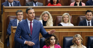 Sánchez cancels his agenda and considers resigning: "I need to stop and reflect"
 The Federal Committee of the PSOE interrupts the event to take to the streets with the militants
 Repsol: "We want to lead generative AI to guarantee its benefits and avoid risks"
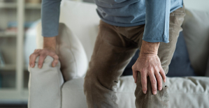 Osteoarthritis: an innovation to improve its management
 Sanofi: demonstration in front of Paris headquarters against job cuts
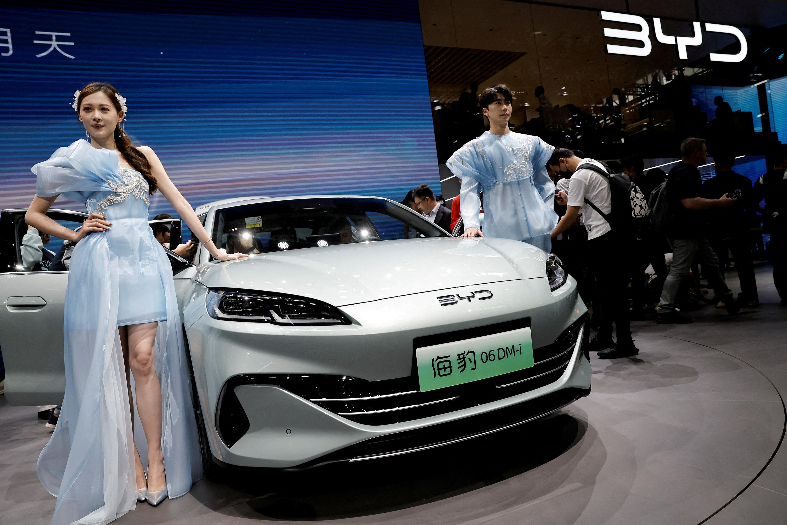
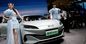 The Chinese car manufacturer BYD sets out to conquer France
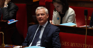 Public finances: after the deputies, Bruno Le Maire asks the senators for savings avenues
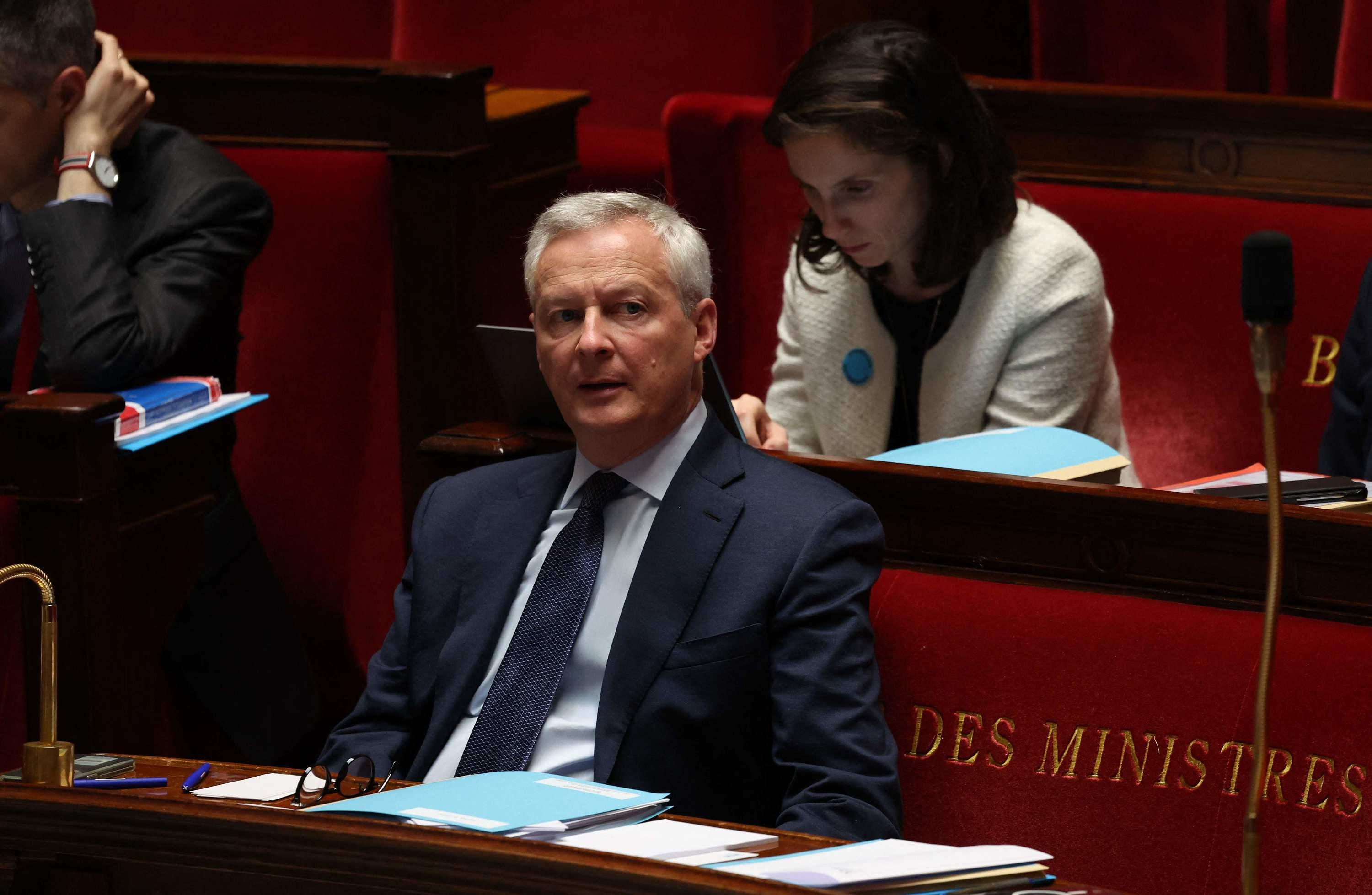
 Faced with opposition from London, a fund supported by Abu Dhabi abandons the purchase of the Daily Telegraph
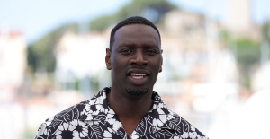 Omar Sy on all cultural fronts
 Jacques Audiard, Swann Arlaud, Benjamin Stora... A hundred men from cinema, theater and books in support of
 Resale, scams and fake tickets: how not to get scammed before Taylor Swift concerts
 Isild Le Besco is not ready to file a complaint against Benoît Jacquot
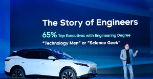 Omoda 7, another Chinese car that could be manufactured in Spain
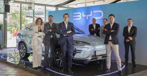 BYD chooses CA Auto Bank as financial partner in Spain
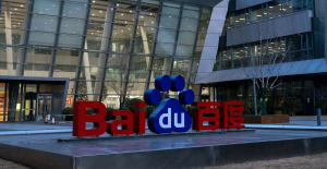 Tesla and Baidu sign key agreement to boost development of autonomous driving
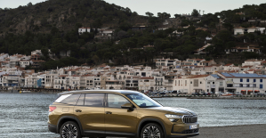 Skoda Kodiaq 2024: a 'beast' plug-in hybrid SUV
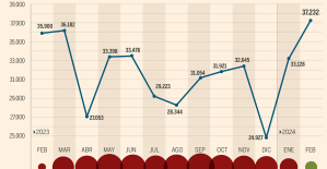 The home mortgage firm rises 3.8% in February and the average interest moderates to 3.33%
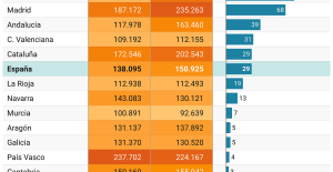 This is how housing prices have changed in Spain in the last decade
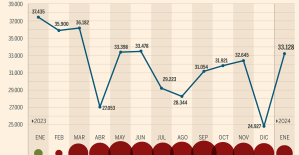 The home mortgage firm drops 10% in January and interest soars to 3.46%
 The jewel of the Rocío de Nagüeles urbanization: a dream villa in Marbella
 Europeans: a senior official on the National Rally list
 Blockade of Sciences Po: the right denounces a “drift”, the government charges the rebels
 Even on a mission for NATO, the Charles-de-Gaulle remains under French control, Lecornu responds to Mélenchon
 “Deadly Europe”, “economic decline”, immigration… What to remember from Emmanuel Macron’s speech at the Sorbonne
 These French cities that will boycott the World Cup in Qatar
 Football: the CAS suspends the sanction of Gabigol who will be able to play again
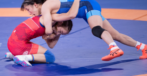 Wrestling: everything you need to know about the sport
 Luis Enrique before Dortmund-PSG: “We must not see pressure as a threat”
 Tennis: the astonishing ploy of Medvedev’s coach to avoid being filmed in his box
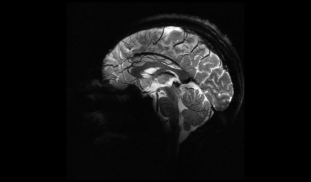
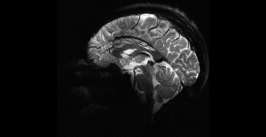
The most powerful MRI in the world has delivered its first images of the human brain near Paris. This extraordinary device, developed by researchers at the Atomic Energy Commission (CEA) on the Saclay plateau (Essonne), is now operational to help better understand the functioning of the brain and certain neurodegenerative or psychiatric diseases.
In 2021, CEA researchers chose to launch the device with a pumpkin, before the health authorities recently gave the green light for the examination of human subjects. Over the last few months, around twenty healthy volunteers have been able to enter the machine's lair, giving rise to the first ultra-precise brain images. “We have a level of finesse never before reached at the CEA,” says Alexandre Vignaud, physicist and research director at the CEA. The magnetic field of this extraordinary magnet reaches 11.7 T (tesla), enough to obtain images ten times more precise than those currently produced in hospitals, where the power of MRI does not exceed 3 tesla.
On Alexandre Vignaud's screen, images of brain sections are compared with what a 3 or 7 Tesla MRI would have given. “With this machine, we can see the very small vessels which supply the cerebral cortex, or details of the cerebellum which were almost invisible until now,” he comments. “Their precision is barely believable!” enthused Research Minister Sylvie Retailleau. “This world first will make it possible to better detect and better treat brain pathologies.”
The machine, a 132-ton magnet housed in a cylinder 5 meters long and as high, made up of a coil carrying a current of 1,500 amps, has an opening of 90 cm to accommodate a human body. This technical feat, the result of a Franco-German partnership, required more than 20 years of research.
Called “Iseult”, the MRI (magnetic resonance imaging device) is the star of Neurospin, the CEA brain imaging research center, directed by neuroscientist Stanislas Dehaene. Two competing projects, in the United States and South Korea, have similar ambitions but have not yet reached the crucial stage of imaging on humans.
One of the objectives of this extraordinary MRI is to refine the understanding of the anatomy of the brain and the areas which are activated when carrying out certain tasks. Scientists already know that different types of images that we are capable of recognizing (a face, a place, a word, etc.) activate distinct regions of the cerebral cortex. With MRI at 11.7 T, “we will be able to better understand the relationship between structure and cognitive functions of the brain, when we read a book or do a mental calculation for example,” assures Nicolas Boulant, director of research at the CEA and scientific manager of the project.
But it will also be a question of elucidating the mechanisms at work in neurodegenerative diseases such as Parkinson's or Alzheimer's, or even in psychiatric conditions (depression, bipolarity, schizophrenia, etc.). “We know, for example, that a particular area – the hippocampus – is involved in Alzheimer’s disease, so we hope to be able to understand the organization and functioning of the cells in this part of the cerebral cortex,” illustrates Anne-Isabelle. Etienvre, director of fundamental research at the CEA.
Researchers also hope to be able to map the distribution of certain drugs, such as lithium, used in the treatment of bipolar disorder. The very high magnetic field of the machine will indeed make it possible to identify the brain structures targeted by lithium in patients and to distinguish more or less good responders to treatment. “If we better understand these very impactful diseases, we should be able to make an earlier diagnosis, and therefore treat them better,” estimates Anne-Isabelle Etienvre.
Iseult will remain dedicated to fundamental research for a number of years. “The device is not intended to become a clinical diagnostic tool, but we hope that the knowledge acquired can then be used in hospitals,” underlines Nicolas Boulant. New healthy volunteers should be recruited by the end of summer. The brains of sick patients will not be studied for a few more years.