Chinese journalist released from prison four years after denouncing China's handling of Covid
 “Russian law” adopted in Georgia: Europeans alongside the streets
 Germany: a new research operation will be launched for a 6-year-old autistic child
 Gaza: under the spotlight, the Israeli-Palestinian conflict shakes up the Eurovision contest
 In Europe, 10,000 people die every day from cardiovascular disease
 Brussels raises Spanish GDP forecasts to 2.1% and foresees a deficit of 3% in 2024
 Inflation rises to 3.3% in April due to gas and food
 “Mediterranean diet” or “DASH”, two good tips for eating better
 Lydia launches Sumeria, an online bank with an interest-bearing account
 The number of Popular Savings Booklets (LEP) is increasing sharply, the “millions of French people” who are entitled to them are called to find out
 INSEE confirms a slight slowdown in inflation, to 2.2% in April
 Paris Olympics 2024: Parisian garbage collectors lift their strike notice after only one day of mobilization
 Death of the great American saxophonist David Sanborn
 Enthoven, Rolin, Sureau... Discover the spring selections of Renaudot
 In support of Palestine, English musicians boycott festivals financed by Barclays Bank
 Where have the sex scenes in cinema gone?
 Omoda 7, another Chinese car that could be manufactured in Spain
 BYD chooses CA Auto Bank as financial partner in Spain
 Tesla and Baidu sign key agreement to boost development of autonomous driving
 Skoda Kodiaq 2024: a 'beast' plug-in hybrid SUV
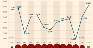 The home mortgage firm rises 3.8% in February and the average interest moderates to 3.33%
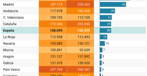 This is how housing prices have changed in Spain in the last decade
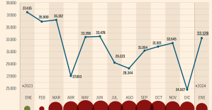 The home mortgage firm drops 10% in January and interest soars to 3.46%
 The jewel of the Rocío de Nagüeles urbanization: a dream villa in Marbella
 Europeans: professor threatened with death Didier Lemaire shows his support for François-Xavier Bellamy
 LFI: Manon Aubry places social issues at the center of her European campaign
 Diving into the secrets of the National Assembly
 Institutions: senators want to restore the accumulation of mandates and put an end to the automatic presence of ex-presidents on the Constitutional Council
 These French cities that will boycott the World Cup in Qatar
 D1 Arkéma: at what time and on which channel to watch the final of the OL-PSG championship
 Nice-PSG, Reims-OM: what is at stake for these late Ligue 1 matches?
 Football: at what time and on which channel to follow Didier Deschamps’ list for Euro 2024?
 Ligue 1: Mbappé and Dembélé absent from the PSG group for the trip to Nice
The photo of his wife Anna Bertha's hand bones together with the ring - made visible by X-rays - is a milestone for very different branches of science. 100 years after the death of Wilhelm Conrad Roentgen, the rays he discovered, with which he examined the hand, are indispensable - and by no means only in medicine. Researchers use them to reconstruct centuries-old murders, viruses can be decoded with high-intensity X-rays, and X-ray telescopes in space reveal high-energy, cosmic processes such as those in black holes.
The discovery more than 127 years ago in Würzburg led to a completely new branch of medicine: radiology. The procedure has helped countless people so far. In 1901, Roentgen received the first Nobel Prize in Physics.
"We have a non-destructive insight into structures," explains Thorsten Bley, Director of the Institute for Diagnostic and Interventional Radiology at the University Hospital in Würzburg, explaining what X-rays can do. “It can be with a mummy, it can also be with a technical device. You can then check whether the connections are intact, whether the metal alloy is tight and has no cracks.”
X-rays are extremely short-wave, high-energy electromagnetic rays that can penetrate and thus shine through many materials. They are not visible to the eye. Bones are easy to see on an X-ray, but soft tissues are not.
Today, taking an X-ray is usually routine, and the radiation dose is much lower than it used to be. "We always do it according to the principle: As little X-ray dose as possible and just as much as necessary," explains Bley, who works with X-rays every day. According to the Federal Office for Radiation Protection, an estimated 130 million X-ray examinations are currently carried out in Germany every year.
Roentgen - born on March 27, 1845 in Lennep, today a district of Remscheid, died on February 10, 1923 in Munich - discovered the rays by accident, late in the evening on November 8, 1895. The scientist experimented in Würzburg with electrical discharges in a pumped almost airless Glass tube (cathode tube).
His laboratory was almost dark at the time. Only the well-known and visible to the naked eye luminous phenomena in the tube illuminated the room weakly. Roentgen wrapped the tube with black cardboard. And watched as a distant fluorescent screen brightened.
What's more: when he held his hand between the tube and the fluorescent screen sometime later – he spent about six weeks almost day and night in the laboratory – Röntgen saw the shadow of his hand bones on the screen. That's roughly how it happened that day, according to the Würzburg Roentgen Board of Trustees.
The association takes care of the physicist's famous place of work with original equipment and devices in Würzburg. Even Roentgen's desk is still in his old laboratory in the university rooms.
Since then, X-rays have revolutionized many areas of research. They have become indispensable in medical diagnostics - in computer tomographs (CT) people are X-rayed slice by slice with them. According to Bley, the latest development in this area is the photon-counting computer tomograph. This provides even more information and enables more precise diagnoses. “This is phenomenal. I'm amazed at the precision every time I see these images.” Bley claims to be working with one of the first 20 of these devices to be installed worldwide.
However, CT devices are not only suitable for examining living subjects. For a study, an international team X-rayed three mummies from pre-Columbian South America that have been kept in European museums since the late 19th century.
The CT scans with the possibility of 3D reconstruction offer unique insights into the body, explains Andreas Nerlich, co-author of the study and head of pathology at the Munich Klinikum Bogenhausen. Formerly, the mummy would have had to be destroyed for such an examination; With conventional X-rays or older CT scans, such a detailed diagnosis is not possible. Result of the work: The researchers were able to show that the people were murdered.
"Aha! Ten minutes of everyday knowledge" is WELT's knowledge podcast. Every Tuesday and Thursday we answer everyday questions from the field of science. Subscribe to the podcast on Spotify, Apple Podcasts, Deezer, Amazon Music, among others, or directly via RSS feed.